Atrial fibrillation
Reducing Stroke Risk with Sensor Technology

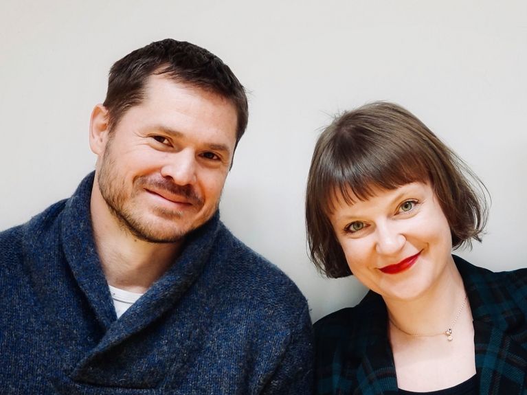
[Translate to Englisch:] Markus Reinthaler und Kate Polak-Kraśna. Foto: Hereon/Hanin Alkhamis, Kate Polak-Kraśna
Atrial fibrillation is one of the most common heart conditions, affecting over 60 million people worldwide. One of its consequences is the formation of blood clots, which can travel to the brain and cause a stroke. Besides blood thinners, a procedure is often used today to seal off the source of these clots. However, the success of this procedure depends on how well the sealing device fits in the heart. The solution could lie in a combination of an adjustable closure device and sensor-guided surgery. Biomedical engineer Katarzyna Polak-Kraśna and physician Markus Reinthaler explain how this works in the interview below.
Image: Hereon
You're currently preparing a spin-off aimed at reducing stroke risk in atrial fibrillation. What is it all about?
Markus: In atrial fibrillation, the contraction patterns of the atria are disrupted and irregular, which slows down blood flow and increases the risk of clot formation. Over the last 20 years, it has been found that 90% of all clots that cause strokes in the brain in these patients form in a small cavity in the left atrium called the left atrial appendage. For those who cannot tolerate the standard blood thinner therapy, there is an option to seal this small outpouching of the left atrium with an occluder. This significantly reduces the risk of stroke.
Kate: The main problem, however, is that this seal doesn’t always fit perfectly. In over 50% of cases, it leaks. Blood clots can then pass by the seal from the atrial appendage, increasing the stroke risk again. That’s why we’ve developed a technology that ensures any potential leaks are immediately detected during implantation, and adjustments can be made to minimize them. This is a promising solution to improve implantation and reduce the risk of stroke.
The Heart with Two Ears
They are called Auricula dextra and Auricula sinistra—right and left atrial appendages. Like small pouches, they protrude into the walls of the atria of our heart. Their exact function is still being researched, but one thing is clear: blood clots can form in them. In the left atrial appendage, atrial fibrillation—a common heart condition—frequently leads to the formation of tiny clots. These clots can then travel to the brain, block a vessel, and cause a stroke. Clots may also form in the right atrial appendage, potentially causing a pulmonary embolism. However, this is medically less significant and likely not relevant.
How does it work?
Markus: To decide where and how to place the seal in the heart, imaging techniques are usually employed, primarily echocardiography. However, the image quality is often not sufficient to confirm the optimal fit of the implant. This is also true for other catheter-based treatment methods. That’s why we are pursuing a sensor-guided approach to improve the implantation of cardiovascular devices. For the atrial appendage closure, our sensors are placed on the surface of the occluder. They allow us to generate real-time information about the position of the implant. This enables the treating physician to see immediately if adjustments are necessary, making the procedure faster, safer, and, of course, more effective.
Is there anything like this already on the market?
Kate: Definitely not. There are no devices with sensor-guided implantation on the market. There is also no real-time feedback directly from the implantation site. As Markus mentioned, the vision during the procedure is provided with imaging techniques. With our sensor, which provides information directly from the implant, we have a unique selling point.
3D-printed occluders. Photo: Hereon/Katarzyna Polak-Kraśna
How did you come up with the idea for the device?
Markus: In cardiovascular medicine, there has been a significant trend in recent years from open surgeries to minimally invasive procedures using catheter technologies. This is also how left atrial appendage closures are performed. When I joined Helmholtz-Zentrum Hereon in 2015, we wanted to work on the next generation of these devices. The current versions only come in standard sizes, which don’t fit every heart perfectly. We explored several approaches and even built some prototypes when Kate joined the team. She brought the idea of sensor technology. We realized that combining our prototype with sensor technology would greatly enhance its application possibilities.
Kate: At that time, more and more reports of leaks during these procedures were appearing in the literature. It was shown that they are quite common, and more data indicated that even small leaks increase stroke risk. We realized that directly addressing these leaks is a crucial approach to making implantation better, safer, and more efficient. The idea of having sensors on-site was then a logical conclusion.
Where are you currently, and what milestones have you set for yourselves?
Markus: So far, we have developed several 3D-printed prototypes to test the mechanical design. We’ve also conducted proof-of-concept studies with the sensors, showing that they can detect the placement in heart tissue. So, the feasibility has been demonstrated. Now we are moving towards creating a prototype that can be used in initial animal trials.
Kate: Exactly. We’ve already shown that our sensors work quite well with animal tissue, but it’s not living tissue. Now we want to ensure that everything works in a living, moving, beating heart. That’s the direction we’re heading. We’re being supported by the GoBio initial program of the German Federal Ministry of Education and Research (BMBF). We’ll begin the project in the coming months. It will take two years, by the end of which we hope to have completed the first animal experiments—and be ready for a spin-off.
Markus, you also work as a physician at Charité. What does the combination of clinical practice and research mean for your project?
Markus: Charité has tremendous expertise in various areas of basic research. Through my work at the German Heart Center of Charité, I maintain good contacts with various research groups. Some of them will now support us in implementing this very ambitious project. Overall, we have tried to bring in experts for all key elements of the project. I think we’ve succeeded quite well in this by collaborating between Charité and Hereon.
Kate: I would say that without Charité and without Markus, this idea wouldn’t even exist. We absolutely need the clinical perspective. Research institutions often find solutions first and then look for the problems. We try to do it the other way around. Thanks to Markus and his colleagues, we get direct insights into clinical practice. This allows us to develop solutions for real problems and work on clinically-driven, needs-based approaches that will have a significant impact on improving healthcare in the future.
The team behind the sensory occluders after the successful pitch for funding from the GoBio Initial programme, from left to right: Adalbert Pakura, Marcin Kraśny, Kate Polak-Kraśna, Markus Reinthaler, Fabian Barbieri. Photo: Hereon/Katarzyna Polak-Kraśna
Kate, what does it mean to you to start a company?
Kate: We are primarily scientists and clinicians. At the beginning, we had absolutely no idea what we were doing. We received funding from the Helmholtz Enterprise Spin-off Program, which allowed us to hire additional team members to develop the sensor technology and marketing strategy. This has enabled us to progress our technology and better understand the entrepreneurial mindset and learn what needs to be done on the path to commercializing our product and starting a business. It has also given us access to many training sessions, coaching, and networking events where we could meet investors and potential partners. So now we have the tools and resources to understand how it works. I enjoy it. Don’t get me wrong—I love being a scientist. But this is something completely new, different, and a very exciting part of my life.
Markus, what is your vision for the future of medicine?
Markus: That’s a very interesting and important question. Because that’s exactly what we’re aiming for: shaping the future. In cardiovascular procedures, which are at the core of our work, there will likely be many more advancements in the coming years. The shift from open-heart surgery to transcatheter procedures will, in my opinion, accelerate. These procedures are becoming increasingly complex. However, to meet this complexity, we need more precise information about the behavior of the implant during the procedure. We aim to address this problem with integrated sensors. With AI-based processing of sensor information along with other data relevant to the procedure, robot-assisted therapy could become a reality in the future. This means that the physician still retains control over the procedure but is supported by assistance systems. This would significantly speed up the learning curve for new procedures.
Kate, do you share this vision?
Kate: Yes, absolutely. I’m glad Markus brought this up. Eliminating the human factor in the clinical context was one of the reasons I wanted to become a biomedical engineer. With sensor technology, we make fewer mistakes because we can see what’s happening inside the patient during the procedure. And we also have the ability to monitor the patient afterwards. We’re continually surprised by how much we still don’t know, learn, and need to understand. The sensors will provide us with the data we need for that. This increases our understanding, and we can continue to improve the procedures. So, I believe that sensor technology has the potential to be a game changer for the future of medicine.
Readers comments